
Brain microstructure modeling from diffusion MRI
Brain microstructure modeling is a crucial topic to provide interpretable measures of the brain usable both to study the normal brain and its changes related to pathologies.This topic of my research considers the modeling and estimattion of brain microstructure from diffusion MRI using advanced multi-compartment models.

Diffusion MRI models processing for clinical studies
While brain microstructure modeling is crucial to understand well the brain, images of complex models need some adapted processing tools to be e.g. registered onto an atlas and analyzed. In addition, diffusion images are prone to artifacts that need to be resolved. This part of my research focuses on these aspects.
Locally most similar atlas construction
High anatomical variability may corrupt atlas-based segmentation results by generating potentially large registration errors. To overcome this drawback, I am interested in the construction of an atlas whose anatomy is as similar as possible to a given patient to delineate. Obtaining efficient methods is also crucial for them to be used in practice.

Locally affine registration
We introduce a new registration algorithm to register in a robust manner predefined anatomical regions. These regions are then registered using a local affine transformation on each region and the global transformation may be computed using the log-euclidean framework on affine transformations. These choices render the algorithm very fast and robust.

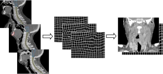
Head and neck atlas construction
Atlas-based segmentation is an efficient and precise way of delineating many structures of a single organ, such as the brain. In this work, we aimed at developing an atlas for segmentation for another region, where cancers are frequent: the head and neck region. To cope with large deformations between the anatomies of the various patients, one must be careful of the registration strategy when building the atlas. We have presented a methodology to create and evaluate a head and neck atlas.

PhD: atlas-based segmentation for radiotherapy
My PhD was about the design and use of anatimocal atlases for the segmentation of brain structures and head and neck structures. These segmentations were then used for the planning of radiotherapy treatment. I introduced several methodological advances also described here, such as locally affine registration, atlas construction from a database, and validation methodologies.